
Mid-South Equine Sports Medicine & Surgery
in Tioga, Texas
Mid-South Equine Sports Medicine & Surgery
in Tioga, Texas
Mid-South Equine Sports Medicine & Surgery provides equine sports medicine, lameness evaluation, and equine surgery for performance horses across North Texas and DFW. We are the only clinic within two hours of Tioga offering standing MRI, traditional MRI, and equine bone scan. Our services include diagnostic imaging, orthopedic and arthroscopic surgery with a board-certified surgeon, regenerative therapy, rehabilitation,gastroscopy, in-house laboratory, pre-purchase exams, and reproduction.
Performance Horse Show Veterinarian | On-Site Care | Mid-South Equine

Equine Diagnostic Imaging
Equine Diagnostic Imaging
North Texas | Tioga, Texas
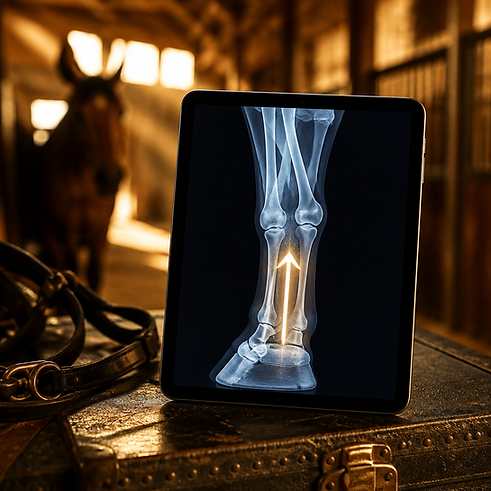
Every imaging modality answers a different question. Radiographs show bone. Ultrasound shows soft tissue, MRI shows both, in detail, inside structures nothing else can reach. Bone scan shows where the skeleton is actively remodeling.
Choosing correctly is most of the work — and it's why we run all four on-site rather than sending horses elsewhere partway through a workup.
Advanced Imaging: Standing MRI & Bone Scan
Mid-South Equine is the only facility within two hours of Tioga offering standing MRI, traditional MRI, and nuclear scintigraphy.
MRI is the next step when a horse blocks sound to a region but radiographs and ultrasound don't explain the lameness —
most often the foot, where the hoof capsule hides the deep digital flexor tendon, collateral ligaments, and navicular bursa from every other modality.
Our standing MRI requires no general anesthesia.Bone scan surveys the entire skeleton at once, identifying active bone remodeling. It's the right tool when the lameness shifts between limbs, won't localize to blocking, or when you suspect pelvic, sacroiliac, or spinal involvement.Full details on standing MRI and bone scan


Digital Radiography
On-site digital radiography produces immediate, high-resolution bone images. We can shoot, review, and adjust the angle in the same session rather than discovering a poorly positioned view after the horse has gone home.
Radiographs are the right tool for:
-
Joint disease and arthritic change
-
Fractures and bone fragments
-
Navicular bone changes
-
Foot balance and hoof-pastern alignment
-
Bone cysts and OCD lesions
-
Pre-purchase survey series
-
Dental and sinus evaluation
What radiographs cannot show is soft tissue. A clean set of foot films does not mean a sound foot — it means the bone looks normal.
That distinction is the single most common source of frustration in lameness cases, and it's why the workup often continues to ultrasound or MRI.
Diagnostic Ultrasound
Ultrasound images tendons, ligaments, and joint structures dynamically — in real time, with the tissue under load.
Common applications:
-
Superficial and deep digital flexor tendon injuries
-
Suspensory ligament desmitis, front and hind
-
Check ligament injuries
-
Joint effusion and synovial evaluation
-
Wound and abscess assessment
-
Reproductive examination and pregnancy monitoring
-
Ultrasound is also how we monitor healing, not just diagnose it.
Soft tissue injuries are re-imaged at intervals through rehabilitation, and the workload advances only when the tissue supports it.
A calendar-based return to work is how good surgical and medical outcomes get undone.

Equine Diagnostic Imaging
North Texas | Tioga, Texas
Every imaging modality answers a different question. Radiographs show bone. Ultrasound shows soft tissue, MRI shows both, in detail, inside structures nothing else can reach. Bone scan shows where the skeleton is actively remodeling.Choosing correctly is most of the work — and it's why we run all four on-site rather than sending horses elsewhere partway through a workup.
In-House LaboratorySame-day bloodwork, chemistry panels, and diagnostic testing performed on-site — results while the horse is still here, not the following week.
Why On-Site Imaging Matters
A lameness workup is a sequence of decisions, and each one depends on what the last image showed. When imaging is outsourced, that sequence breaks into appointments spread over weeks, with a different person interpreting each step.Here, blocking, imaging, and interpretation happen in one visit. If the radiographs are clean and the horse blocked to the foot, we can go to MRI the same day rather than scheduling you back in three weeks.
-
Standing MRI
-
Traditional MRI
-
Digital radiography
-
Ultrasound imaging
-
Nuclear scintigraphy